

Preview
Identifier
Winner, Honorable Mention
Creation Date
Spring 2020
Department
Biological Sciences
Narrative
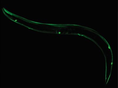
My research in Professor Vidal-Gadea's lab has consisted of studying which versions of the protein dystrophin are present during exercise in the microscopic worm, C. elegans. This image depicts green fluorescence seen when the long version of dystrophin is expressed. To create this image, we injected a small piece of DNA, our fusion product, into a C. elegans. The fusion product consists of a promoter region of the long version of dystrophin connected to green fluorescent protein (GFP) and an unc-54 3’ untranslated region (UTR). The promoter controls where and when the fluorescence is expressed, and the UTR tells the animal to make a lot of fluorescence. So, everything green in the image is where the long version of dystrophin is located. The next step in our research is to inject the same worm with the short isoform dystrophin promoter, which will show up red in the image. With this data, we will be able to compare red to green fluorescence, which will tell us the ratio of short to long dystrophins being expressed.